


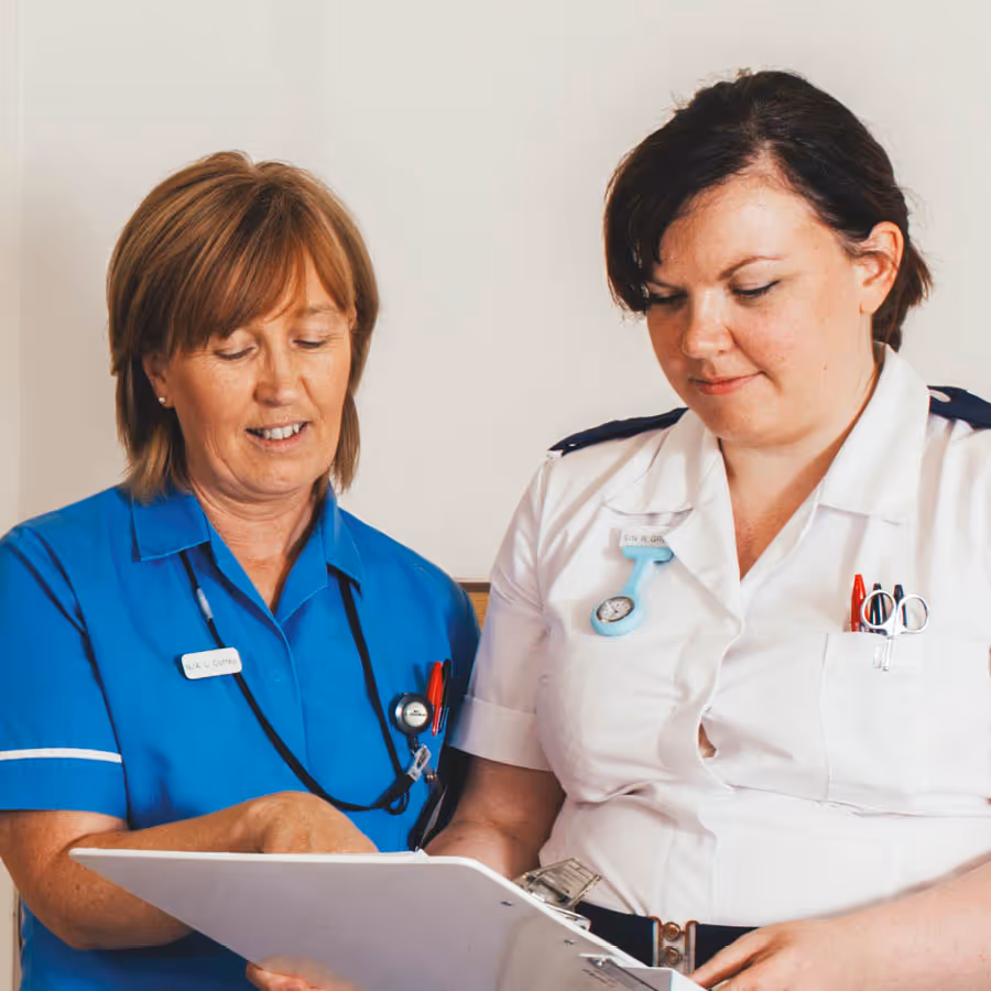
An echocardiogram, also known as an echo, is a non-invasive imaging test used to examine the heart and is performed by a cardiac physiologist.
How it works
- A small probe is placed on the chest wall with ultrasound gel. Echoes are captured by the probe and transformed into a moving image displayed on a monitor during the scan.
An echocardiogram helps diagnose and monitor various heart conditions by assessing
- Heart Structure: It examines the heart’s anatomy, including its chambers, valves, and blood vessels.
- Blood Flow: It analyses how blood flows through the heart.
- Pumping Chambers: It evaluates the function of the heart’s pumping chambers.
Common uses of echocardiograms include detecting
- Damage from a Heart Attack: Identifying areas affected by reduced blood supply.
- Heart Failure: Assessing the heart’s ability to pump blood effectively.
- Congenital Heart Disease: Detecting birth defects affecting heart function.
- Heart Valve Problems: Evaluating valve function and blood flow.
- Cardiomyopathy: Assessing thickening or enlargement of heart walls.
- Endocarditis: Detecting infections that damage heart valves.
Process
You’ll be asked to remove any clothing covering your upper half before lying down on a bed. You may be offered a hospital gown to cover yourself during the test.
When you’re lying down, several small sticky sensors called electrodes will be attached to your chest. These will be connected to a machine that monitors your heart rhythm during the test.
An ultrasound gel will be applied to your chest or directly to the ultrasound probe. You’ll be asked to lie on your left side and the probe will be moved across your chest.
The probe is attached by a cable to a nearby machine that will display and record the images produced.
You won’t hear the sound waves produced by the probe, but you may hear a swishing noise during the scan. This is normal and is just the sound of the bloodflow through your heart being picked up by the probe.
The whole procedure will usually take between 30 and 60 minutes, and you’ll normally be able to go home shortly afterwards.
No Preparation needed.
LEAFLETS - Information for patients
No items found.


Our team
Lorem ipsum dolor sit amet, consectetur adipiscing elit, sed do eiusmod tempor incididunt ut labore et dolore magna aliqua.


